

LIVE WEBINAR SERIES
Cherenkov Imaging
in Radiation Therapy
Thursdays, November 5th, 12th and 19th
12-1pm EST
Receive continuing education credits. See below for details.Join Dartmouth Health and DoseOptics for a 3-part webinar series exploring current and emerging applications of Cherenkov imaging in clinical radiation therapy.
Developed by a multidisciplinary team of physicians, medical physicists and engineers, the series brings together leading researchers, clinical experts and recent adopters. Through focused presentations and a moderated Q&A, attendees will learn how Cherenkov imaging is being used to enhance treatment delivery verification, support patient safety and provide additional insight during radiation therapy.



Hosted byReceive Continuing Education Credits Accreditation: In support of improving patient care, Dartmouth Health is jointly accredited by the Accreditation Council for Continuing Medical Education (ACCME), the Accreditation Council for Pharmacy Education (ACPE), and the American Nurses Credentialing Center (ANCC), to provide continuing education for the healthcare team. American Medical Association (AMA) Dartmouth Health designates this live activity for a maximum of 3.0 AMA PRA Category 1 Credit(s)™. Physicians should claim only the credit commensurate with the extent of their participation in the activity. Commission on Accreditation of Medical Physics Education Programs (CAMPEP)
An application has been submitted to CAMPEP for approval of 0.75 MPCEC hours per webinar.
CATCH. VERIFY. CORRECT.

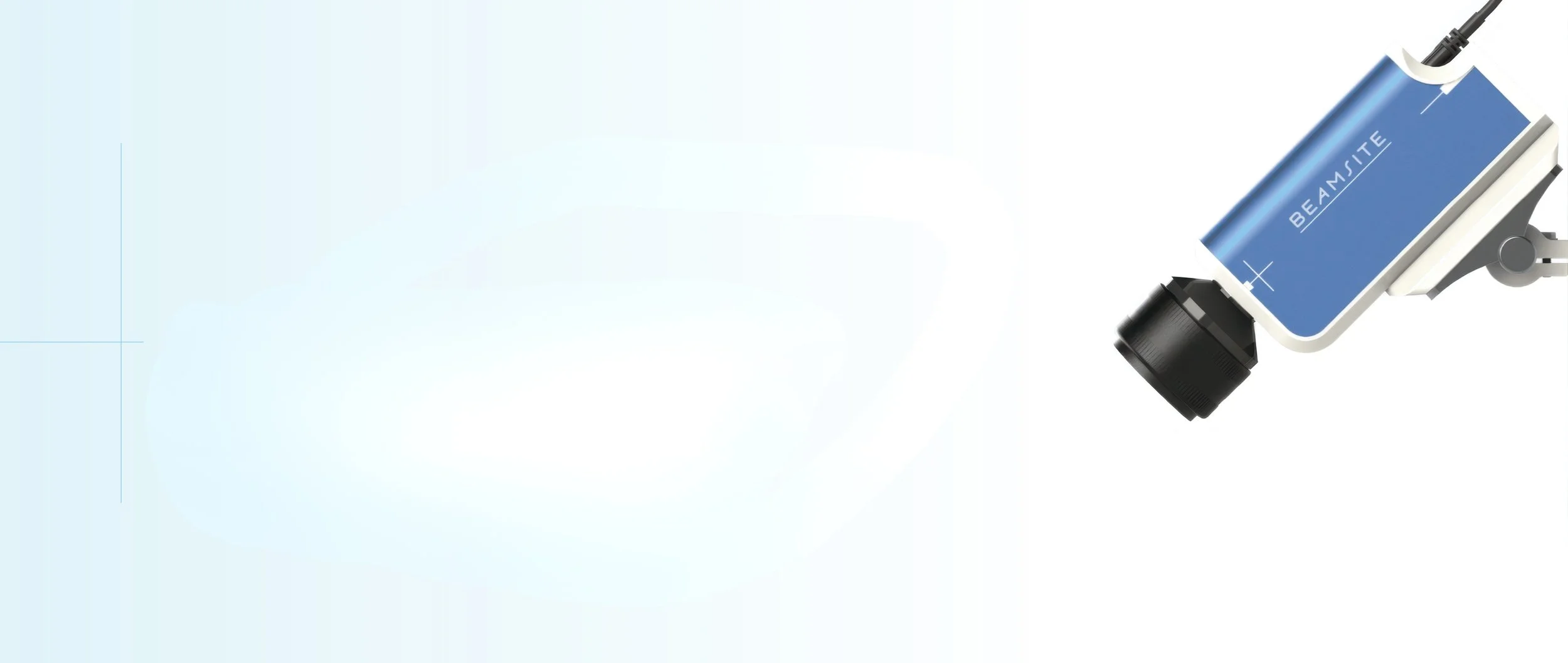
BeamSite® captures Cherenkov light emitted during radiation therapy to provide real-time visualization of radiation directly on the patient without adding ionizing dose, setup time, or workflow burden.
Reveal hidden errors and support upstream clinical improvements with BeamSite. By helping the treatment team verify dose delivery in real time, BeamSite provides meaningful insight even in the face of patient variability or differences in imaging workflows.
By enabling true, real-time treatment monitoring, it bridges the gap between physics and clinical workflow, helping teams uncover trends, improve consistency, and elevate overall treatment quality.
See What Others Can’t
Always on. No additional setup required.
Improves safety with real-time visual feedback.⁴
Visualizes both entry and exit beams.
Helps identify and correct issues as part of routine QA.
Offers multi-camera support for full-body coverage.
Records treatment sessions to help identify anomalies for later review with your team.
BeamSite in the Clinic
The patient images and videos below were captured from BeamSite cameras installed in three LINAC bunkers throughout the Radiation Oncology Department. These details highlight some of the incidents identified during patient treatments; for more information, please see the full manuscripts referenced below.
With BeamSite, your clinic can streamline operations, optimize treatment outcomes, guarantee your patients’ peace of mind–and save lives!


1. Not FDA cleared for electrons, protons or brachytherapy2. Alexander, Daniel A. et al. “Retrospective Evaluation of an Always-on Cherenkov Imaging System for Radiotherapy Quality Improvement.” (2021, preprint available online: http://arxiv.org/abs/2110.07494).3. Jarvis, Lesley A et al. “Initial Clinical Experience of Cherenkov Imaging in External Beam Radiation Therapy Identifies Opportunities to Improve Treatment Delivery.” International journal of radiation oncology, biology, physics vol. 109,5 (2021): 1627-1637. doi:10.1016/j.ijrobp.2020.11.0134. Robinson A, Tallhamer M, Stieler F. A Review of Cherenkov Imaging for Real Time Verification in Radiation Therapy. Pract Radiat Oncol. 2026Case Study
Vel volutpat ac sed turpis porttitor varius. In ante cras scelerisque placerat penatibus posuere tellus velit pellentesque.
Pellentesque dui felis mattis pretium amet vestibulum sed aenean venenatis. Tincidunt blandit pulvinar vel auctor consequat amet velit. Condimentum amet nec dui malesuada arcu egestas feugiat. Vitae massa et ipsum congue mattis. Justo ornare viverra neque amet sapien odio habitasse mi quisque. Odio egestas neque urna sed at enim amet pellentesque.

FAQs
-
When tissue is irradiated with high energy photons (or electrons) during radiotherapy, it will glow with visible light due to the Cherenkov effect. Although extremely dim, this light can be seen with BeamSite, which has been designed specifically to image Cherenkov light in radiotherapy treatment rooms.
-
Yes! BeamSite doesn’t contribute any additional radiation to a patient’s treatment–it is simply a camera system that allows healthcare professionals to visualize and monitor what’s happening. The imaging produced is “free” information as a result of a patient’s treatment.
-
Yes. BeamSite offers real-time monitoring and is cleared for use only with photon external beam radiotherapy, anywhere on a patient’s body.
-
Yes. BeamSite works by imaging synchronously with the linear accelerator, thereby minimizing the impact of room light.
-
Yes. BeamSite has FDA 510(k) clearance for sale in the US.
-
BeamSite requires direct line of sight to the tissue it is imaging. Sheets or clothing over the tissue being treated may block some of the light coming from the patient.
-
There are more than 50 academic publications on Cherenkov imaging in radiotherapy. See a selected list here.
-
Not currently. BeamSite relies on the clinical experience of the Radiation Therapist to identify when the treatment does not look appropriate.
Request a Meeting
Ready to see the power of BeamSite in action? We’d love to connect with you, show you how it works, and answer any questions you may have.