
Beyond the Treatment Plan: Monitoring Contralateral Breast Dose in Real Time
Radiation therapy is highly effective for treating breast cancer, but minimizing unnecessary radiation exposure to healthy tissue remains an important clinical objective. One area of particular concern is unexpected dose to the contralateral breast, especially in younger patients.
Research from the Women’s Environmental, Cancer, and Radiation Epidemiology (WECARE) study demonstrated that women diagnosed with breast cancer before age 40 who received more than 1 Gy of radiation to the contralateral breast had approximately a 2.5-fold increased risk of developing a second primary breast cancer in the opposite breast. While the absolute risk remains relatively low, younger patients have both greater tissue radiosensitivity and a longer life expectancy during which radiation-induced malignancies may develop.
Treatment techniques such as deep inspiration breath hold (DIBH), image guidance, and advanced treatment planning have significantly reduced exposure to healthy tissues. However, variations in patient positioning, anatomy, or motion can still result in unintended radiation reaching areas outside the intended treatment field.
This is where real-time treatment visualization provides additional clinical value. BeamSite® uses Cherenkov imaging to visualize radiation delivery directly on the patient's surface during treatment. By providing real-time feedback, it helps clinicians confirm treatment accuracy and catch radiation in unexpected areas.
While the strongest WECARE evidence is for women under 40, many clinicians and researchers often extend the principle to patients under 50, since younger age at treatment remains a recognized risk factor for radiation-induced secondary cancers.
51-year-old female undergoing 3D conformal treatment with DIBH to her left breast.¹
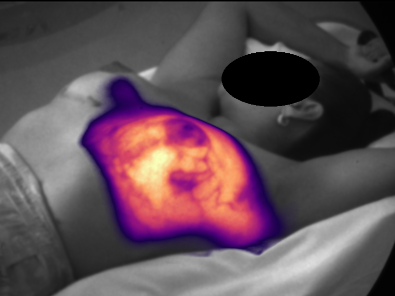
BeamSite displays unexpected contralateral breast dose during fraction 5 of treatment.¹
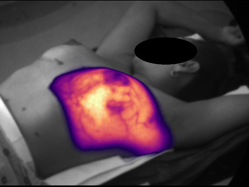
DIBH thresholds were tightened based on BeamSite
imaging observations, reducing contralateral breast
dose in future treatment fractions.¹
In one breast radiotherapy case, BeamSite revealed unexpected dose to the contralateral breast despite a patient remaining within established DIBH thresholds. The clinical team subsequently tightened the DIBH parameters, reducing contralateral breast dose in future treatment fractions.
BeamSite provides an additional layer of treatment verification. For younger breast cancer patients, where minimizing contralateral breast dose is particularly important, real-time visualization may help identify opportunities to further refine treatment delivery and support continuous quality improvement.
1. Robinson A, Tallhamer M, Stieler F. A Review of Cherenkov Imaging for Real-Time Verification in Radiation Therapy. Pract Radiat Oncol. 2026